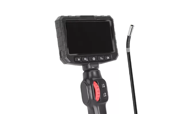
Preparation:
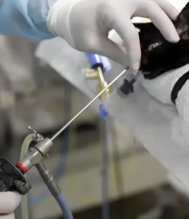
Administer appropriate anesthesia to ensure pet immobility.
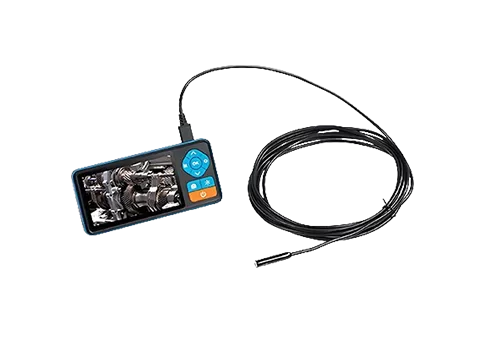
Connect the gastroscope to the video processor and test the LED light source for clarity.
Device Insertion:
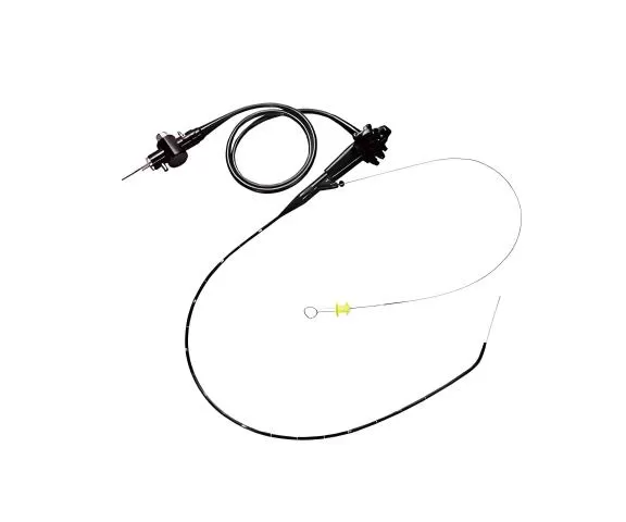
Gently introduce the flexible shaft (diameter 5–12mm) transorally into the esophagus.
Advance slowly through the esophageal sphincter while monitoring resistance.
Real-time Visualization:
Observe the 1080p endoscopic feed for gastric mucosa, ulcers, or foreign bodies.
Document hyperemia, erosions, or tumors via high-resolution imaging.
Diagnosis & Documentation:
Analyze findings (e.g., Helicobacter infections, neoplasia).
Capture biopsy samples and generate annotated digital reports.
