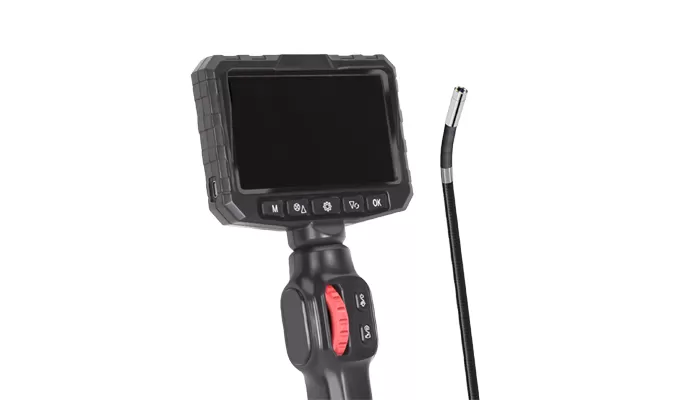
Veterinary Flexible Laryngoscope Clinical Procedure Steps
Preparation:
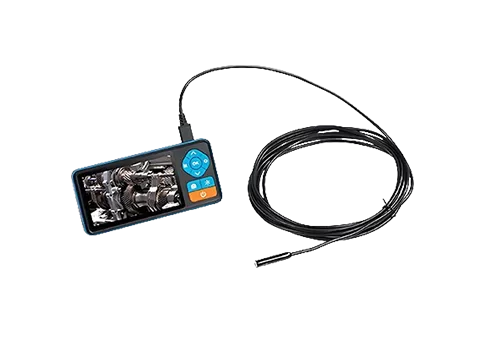
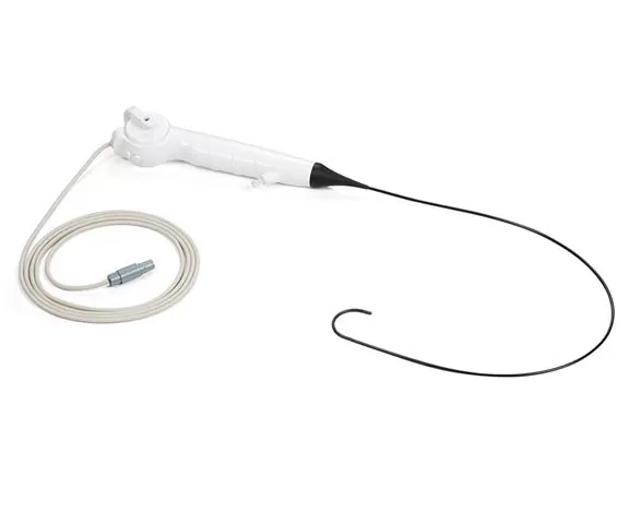
Power on the external display system and connect the flexible tubular probe to the handle.
Adjust LED illumination for optimal visibility in the oropharyngeal cavity.
Device Insertion:
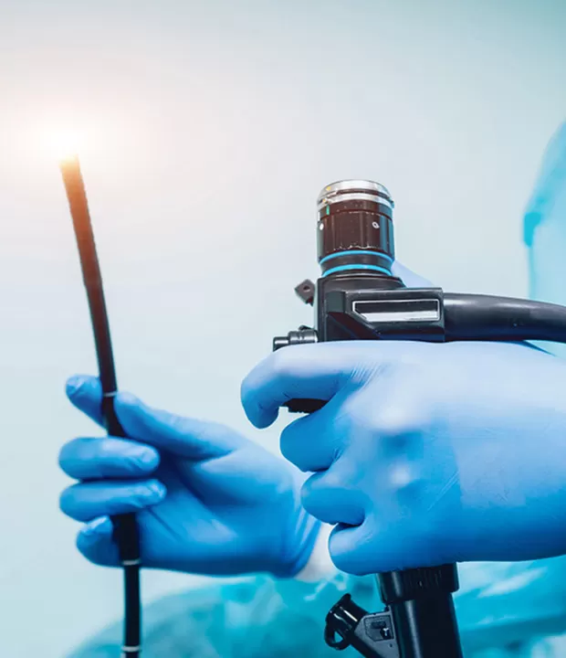
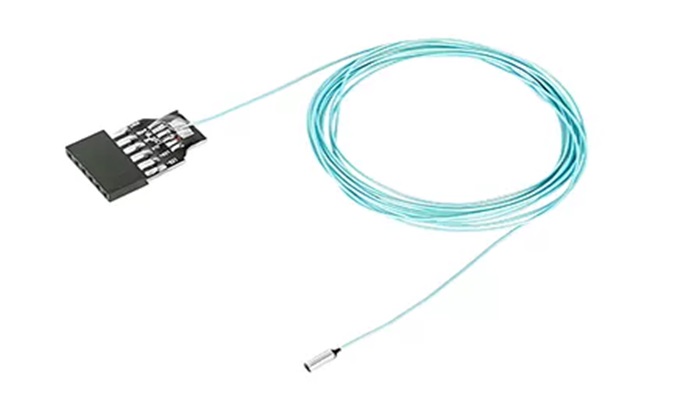
Gently insert the flexible probe (diameter 4–8mm) transorally with the pet's neck extended.
Navigate past the tongue base using ergonomic handle articulation.
Real-time Visualization:
Monitor the external display for live video of vocal cords and glottis.
Capture images/video of lesions, edema, or foreign bodies via the external system.
Diagnosis & Documentation:
Analyze visual findings (e.g., polyps, inflammation, tumors) on the external display.
Annotate abnormalities and generate electronic medical reports through the connected interface.
