Preparation:
Sedate the pet and position for optimal airway access.
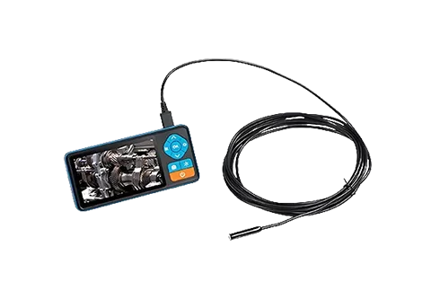
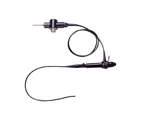
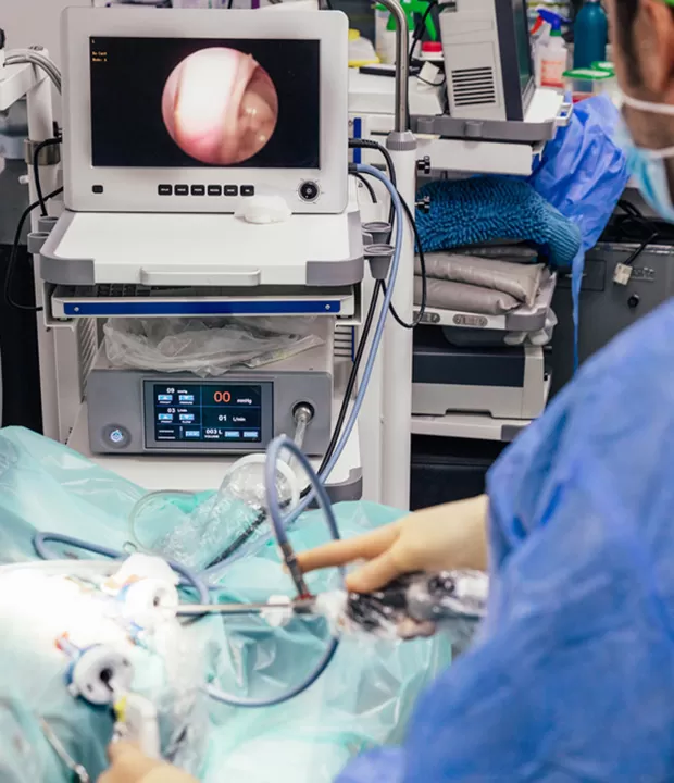
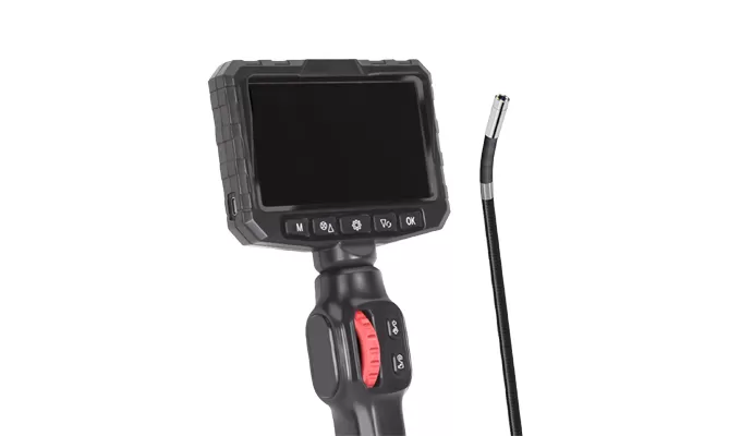
Connect the bronchoscope to the light source and external monitor.
Device Insertion:
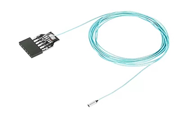
Gently advance the scope transorally/nasally through the trachea.
Navigate bronchial branches using ergonomic controls.
Real-time Visualization:
Monitor airways for inflammation, masses, or foreign bodies on-screen.
Capture images/video of pathology via external system.
Diagnosis & Sampling:
Assess lesions (e.g., granulomas, bleeding) and perform BAL/biopsy if needed.
Document findings and generate electronic records.
